Table of Contents
Detectors
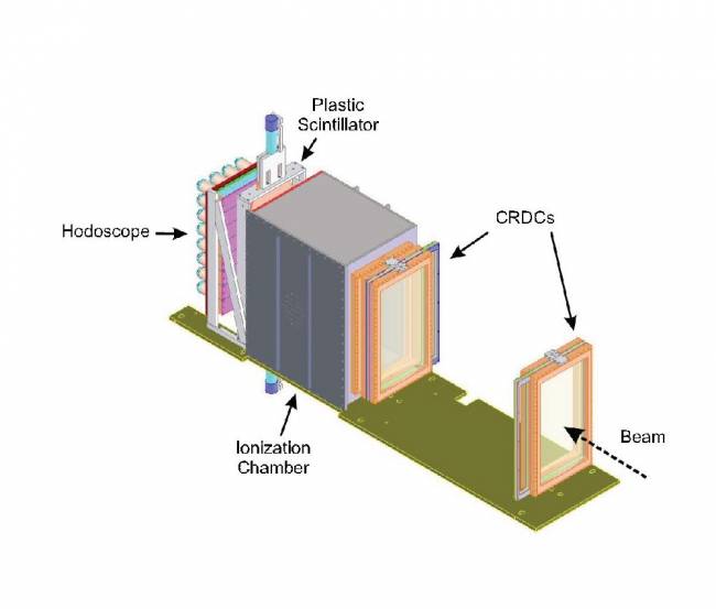
The standard detection system of the S800 consists of a plastic scintillator at the object station; two tracking detectors at the intermediate plane station (TPPAC) and a series of detectors at the focal plane station (see figure below), which include two cathode readout drift chambers (CRDC) located about 1 m apart; a multi-segmented ionization chamber, a thin plastic scintillators and a Hodoscope.
Plastic scintillators
In order to determine the Time-Of-Flight for the particle identification, the S800 includes a plastic scintillator at the object station (S800_OBJ) and at the focal-plane station (E1). The detector material typically used is BC-400 or BC-404 made from polyvinyltoluene (>97% ) and organic fluors (<3%) with a density 1.032 g/cm3 and a refractive index 1.58. The thickness of the detectors is chosen on the basis of the charge of the nuclei to be measured. The available thicknesses are 127 μm and 1 mm for OBJ_SCI and 1 mm and 5 mm for E1. (Guidelines explaining the installation of E1 scintillator can be found here). The OBJ_SCI has an active area of xxx and is connected to a photomultiplier assembly H6533. The E1 scintillator is connected to photomultipliers EMI 98807B in both ends (up and down). The time signal from the E1 scintillator is calculated as the average time signal from each photomultipliers. Different Time-of-flights can be constructed by combining the timing signals from these two detectors with the timing signals from the A1900 focal plane, and the RF cyclotron. The E1 detector is also used to define a valid trigger from the S800. The timing resolution for a point-like beam spot in the focal plane is around 100 ps. However, this resolution worsens significantly (up to 1 ns) when the whole focal plane is illuminated, because of path length differences of the traversing nuclei. It can be recovered by tracking the position of each event on the scintillator from the position and angle information provided by the CRDC detectors. The plastic scintillators can withstand maximum rates up to 1 x 10⁶ particles per second.
Tracking Parallel Plate Avalanche Counters (TPPAC)
Some experiments are particularly sensitive to the incoming positions and angles of the nuclei impinging on the target. Two tracking parallel plate avalanche counters (TPPAC) are installed in the intermediate plane station of the analysis line. The position and angles measured with both TPPACs are transformed into the corresponding coordinates in front of the target, using the transfer matrix of the second half of the analysis line. The analysis line dipole magnets downstream of the intermediate image plane filter the particles produced in the tracking detectors, which would otherwise contaminate the data.
Each TPPAC has an active area of 10 cm x 10 cm and is filled with isobutane at a typical pressure of 7 torr. The detector consists of a cathode foil with a series of aluminum strips oriented in the non-dispersive direction, followed by an anode plate and a second cathode foil with the strips oriented in the dispersive direction. A total of 128 pads are connected to the strips of each cathode foil. The x and y positions are determined from the charge distribution on the pads. The position calibration was done using the pad pitch of 1.27 mm.
The particles transmitted through the TPPAC ionize the gas, producing electrons and positive ions. The drift of electrons towards the central anode plane induces an image charge on the aluminum strips. The signal generated on a given pad is sent to a preamplifier, and processed by a switch capacitor array (SCA), which acts as an analogic memory. Since the signals of this detector are generated before a valid trigger occurs, they need to be temporarily recorded until the trigger is received. The SCA samples the signals from the detector with a period of 200 ns and saves the data on a continuous mode, generating an analogic buffer. When a valid trigger is received, the sampling stops and the SCA pointer moves back in the buffer by a number of samplings pre-defined according to the time passed between the tracking signal and the valid trigger. In this way, the valid trigger is correlated with the tracking signal corresponding to the same event. The reading algorithm is lead by the FPGA chips of a XLM72 VME module. The TPPACs can work at maximum rates is of order of 3 x 10⁵ particles per second. The efficiency is significantly reduced for light nuclei (typically below Z=10).
Cathode Readout Drift Chambers (CRDC)
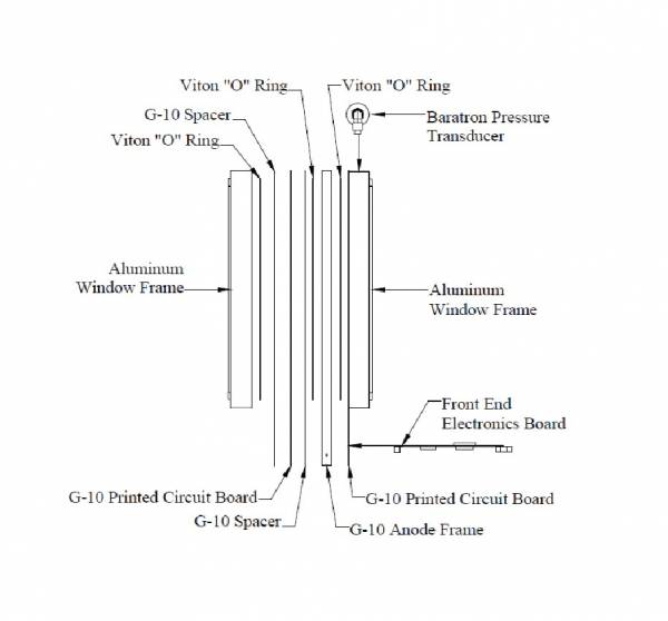
Two Cathode Readout Drift Chamber (CRDC) are used to measure the transversal positions and angles in the focal plane. The first detector (CRDC1) is located at the nominal optical focal plane, and it is separated 1 m from the second downstream detector (CRDC2). Each detector has an active depth of 1.5 cm, an active area of 26 cm (non-dispersive direction) x 56 cm (dispersive direction), and it is filled with a gas mixture consisting of 80% CF4 and 20% C4H10 at a typical pressure of 40 torr. The detector frame has a volume of 68 cm (dispersive) x 38 cm (non-dispersive) x 10.3 cm (depth). The operating high power depends on the charge of the measured nuclei. A schematic view of a CRDC can be seen in the figure below.
Each detector consists of two 12-µm PPTA windows mounted on frames, two printed circuit boards (PCB) and an anode frame. Each PCB is made of un-masked G-10, and includes a field shaping foil (70 µg/cm2 polypropylene with 0.1 µm of evaporated gold) to ensure a uniform field in the active region of the detector. Two G-10 spacers are laminated to the board on each side. The shaping foils are made of 1.9-mm pitch evaporated aluminum strips perpendicularly oriented to the electric field. The anode frame includes a glued cathode grounding plane, an anode wire running across the field, and a Frisch grid. Cathode pads are located in front of and behind the anode wire. The pads have a pitch of 2.54 mm. The anode frame is sandwiched between the two printed circuit boards with two spacers in between, as shown in the figure below.
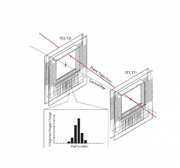
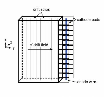
The principle of operation of a CRDC is illustrated in the figure below. The nuclei passing through the detector ionize the gas, dissociating electrons which drift towards an anode wire under the action of a electric field. The collection of charge in the anode induces a positive charge on the cathode pads, which are read out individually. The x position (dispersive direction) is determined by fitting the charge distribution on the cathode pads with a Gaussian function. The drift time of the electrons to the anode wire, measured with respect to a trigger signal (typically from the E1 scintillator), provides the y position (non-dispersive direction). The resulting position resolution is less than 0.5 cm. Depending on the position of the track, the typical drift times of the electrons to the anode wires are 0 to 20 µs. The relatively long drift times limit the maximum rate that the detector can process properly to around 5000 counts per second. High rates affect also the aging of the anode wire. These problems can be partly amended by spreading the beam over a large portion of the active area, as it is done in focus mode. In addition, an electrostatic gate has been added above the Frisch grid of the CRDCs to enable the possibility to block primary electrons based on a trigger decision. This functionality is still under development, regarding in particular the recovery time of the preamplifiers after switching of the electrostatic gate. If successful, it should mitigate the effects of high rate on the CRDCs, by effectively reducing the rate of avalanches on the wire. Note that it will not eliminate pile-up effects, which can only be reduced by increasing the drift voltage (standard operating anode voltage: 800 V, maximum operating voltage: 1000 V; nominal operating cathode voltage: -1000 V, maximum operating voltage: -1300 V).
Both CRDCs are equipped with digital electronics, which consist of seven front-end electronic boards (FEE) designed and developed by the STAR collaboration (RHIC), followed by interface boards connected to a programmable FPGA VME module (XLM72) like the one used with the TPPACs in the intermediate image station. Each FEE includes 32 channels of preamplifier shaper, followed by a switch capacitor array (SCA) and an ADC. The processing of signals is driven by the FPGA module. Each SCA samples the signals after a valid trigger is received and sends the information into the ADC. The digitized data are then stored into the internal memory of the FPGA and read out in block mode. The sampling frequency and number of samples read out are adjustable; typical values are 20 MHz and 8 to 12 samples. The time needed for each sampling is around 16 µs. Thus, the dead time of the electronics is directly proportional to the number of samples read out. The main advantage of the on-detector digitalization technique used with the CRDCs is the reduction of noise by avoiding the transmission of analog signals (448 from the two CRDCs) outside the vacuum chamber, and the possibility to record multi-hit events like in traditional TPC detectors. The schematic diagram of the firmware for the reading of the XLM72V FPGA can be found here.
Ionization chamber
An ionization chamber downstream of both CRDCs is used to identify the Z number of the transmitted nuclei from their energy loss. The detector has an active area of approximately 30 cm x 60 cm and a depth of approximately 406 mm (16 inches). It is filled with P10 gas (90% argon, 10% methane) at a typical pressure of 300 torr, although this value can be increased up to 600 torr for light nuclei. The detector consists of 16 stacked-parallel plate ion chambers with narrow anode-cathode gaps, placed along the detector’s central axis, perpendicular to the beam direction (see figure). The plates of each of these stacked chambers are constructed from 70 µg/cm2 polypropylene with 0.15 µm of aluminum evaporated on each side. The entrance and exit windows of the chamber are made of 12 mg/cm2 Mylar with an overlay of Kevlar filaments and epoxy.
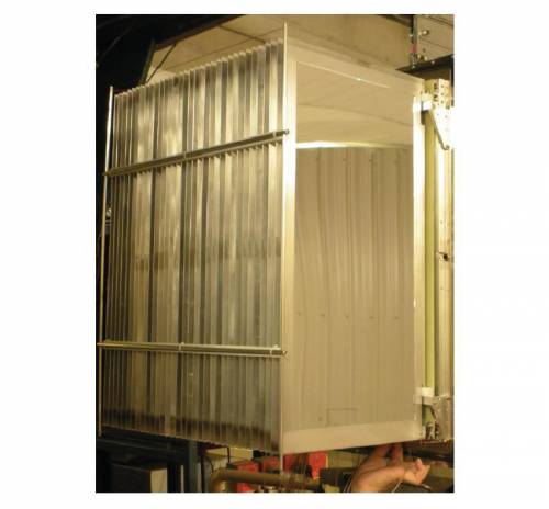
The electrons and positive ions liberated by the ionization of the gas along the particle trajectory drift towards the closest anode-cathode pair. The drifting electrons and ions absorb the energy stored in the detector capacity and produce a voltage change of the anodes across the resistor. The main advantages of the anode-cathode configuration is that the electrons and ions are collected on a very short distance (about 1.5 cm), thus reducing pile-up and position dependence of the signals. Moreover, dividing the detector into 16 sections reduces the detector capacitance and consequently its noise. Each anode is attached to a small preamplifier inside the ion chamber. This significantly reduces the electronic noise, although it involves the venting of the whole chamber whenever a malfunctioning preamplifier needs to be replaced. The electronic signals from the preamplifier are sent into a CAEN N568B 16-channel shaper/amplifier with remotely adjustable gains. The output signals feed a Phillips 7164H ADC. The energy-loss resolution of the ionization chamber can be significantly improved after correcting the position and momentum dependences. Elements up to Z=50 can be separated.
Hodoscope
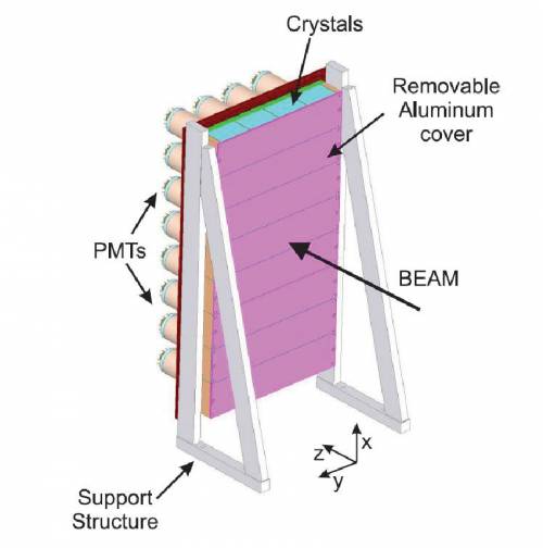
A CsI(Na) hodoscope detector located downstream of the E1 scintillator is used to measure the total kinetic energy of implanted nuclei, allowing the identification of different charge states. An additional use recently tested is the measurement of isomer gamma-rays emitted from implanted nuclei.
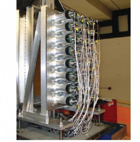
The hodoscope is composed 32 sodium-doped cesium iodide CsI(Na) scintillating crystals manufactured by ScintiTech. Each crystal is 5.1 cm-thick, has an active area of 7.6 cm x 7.6 cm, and is attached to a photomultiplier (Hamamatsu R1307). The photo-cathodes are made of a bi-alkali material with a transmission peak at 420 nm. The 32 crystals are arranged in eight rows of 4 crystals each so as to cover approximately the same solid angle as the CRDCs. The frontal and lateral sides of each crystal are covered with two 150-µm thick layers of a white Teflon reflective material to provide light shielding between the crystals. The photocathodes are connected to a CAEN N568B 16-channel shaper/amplifier, followed by a Phillips 7164H 12-bit ADC. The signals from the crystals are gain-matched to a middle position in the ADC spectra by varying the biases of each photocathode.

